772–774 ECCLESALL ROAD  ·  SHEFFIELD  ·  S11 8TB   info@sheffieldveinclinic.co.uk

Case Study · Sheffield Vein Clinic

When a Varicose Vein Turns Angry: A Case of Superficial Thrombophlebitis

A hard, red, painful lump appearing on a varicose vein is easy to mistake for a bruise. It usually isn’t one — and it’s worth getting checked properly. Here’s a real case from our clinic, from the first scan through to full recovery.

Condition
Superficial thrombophlebitis
Underlying cause
Left great saphenous vein reflux
Scan
Duplex ultrasound
Treatment
Full-length EVLA + foam sclerotherapy
Clinician
Professor Stephen Goode, Consultant Interventional Radiologist

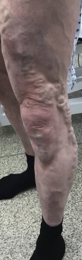
This patient came to us with a long history of varicose veins in the left leg, caused by underlying reflux in the great saphenous vein (GSV) — the main superficial vein running up the inside of the leg. Over the space of a few days, a section of one of the varicose tributaries had become red, hard, and painful to the touch, with a brownish discolouration spreading along the skin above it. This combination — pain, redness, and a lump you can feel tracking a vein — is what we call superficial thrombophlebitis, and it’s more common than people realise.

What is superficial thrombophlebitis?

It sounds alarming, but broken down it simply means inflammation (“-itis”) of a superficial vein (“phlebo-”) caused by a small blood clot forming inside it (“thrombo-”). It happens most often in a vein that’s already varicose, because slow, turbulent blood flow through a stretched, weakened vein makes clotting more likely. The result is a vein that goes from being a cosmetic nuisance to something genuinely sore, red, and hard almost overnight.

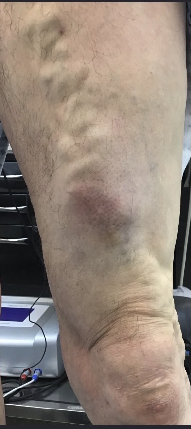
Inflamed, thrombophlebitic superficial varicose vein on the lower leg before treatment, showing redness and discolouration tracking the vein
Fig. 1 — The affected leg at first assessment. Alongside the network of varicose veins, there is a clearly defined patch of redness and discolouration over the mid-calf — the site of the thrombophlebitis.

It’s a fair question to ask: is this actually dangerous? The honest answer is that superficial thrombophlebitis on its own usually isn’t — but it can be a warning sign, because in a small number of cases the clot can extend towards the deeper veins in the leg, where the risks are higher. That’s exactly why we don’t rely on how it looks or feels alone. Every case like this gets a duplex ultrasound scan.

Is this the same as a DVT?

No — and the difference matters. A deep vein thrombosis (DVT) sits in the deep venous system, doesn’t show on the skin, and carries a meaningfully higher risk of serious complications. Superficial thrombophlebitis, by contrast, is a clot in a vein close to the surface, which is why it shows up as visible redness and a firm, tender lump. On its own, it’s generally the less risky of the two.

That said, “less risky” doesn’t mean “no risk.” A clot in a superficial vein can, in a minority of cases, propagate further along the vein, or a fragment can dislodge and travel into the deep venous system. From there, in rare cases, it can migrate further and cause a pulmonary embolism (PE) — a clot in the lungs. The scenario we’re specifically watching for is a clot that fills the entire great saphenous vein and reaches the sapheno-femoral junction (SFJ), where the superficial and deep systems meet. This is the highest-risk pattern, because the clot then sits right at the doorway to the deep system. It’s precisely this risk that makes duplex ultrasound essential rather than optional, and why the scan findings — not just the clinical appearance — drive the treatment decision.
Signs and symptoms to watch for

Superficial thrombophlebitis usually announces itself fairly clearly, but it’s worth knowing exactly what to look and feel for along the line of a varicose vein:

  • A firm, rope-like or cord-like lump you can feel under the skin, following the path of a vein
  • Redness or a darker, brownish discolouration of the skin directly over the vein
  • Tenderness, aching, or pain when the area is touched or pressed
  • Warmth over the affected area compared with the surrounding skin
  • Mild localised swelling, usually confined to the immediate area rather than the whole leg
Red flags — seek urgent medical attention

Go to A&E urgently if you develop significant swelling of the whole leg, chest pain, shortness of breath, or coughing up blood. These can be signs that a clot has spread to the deep veins or the lungs, and need immediate assessment.

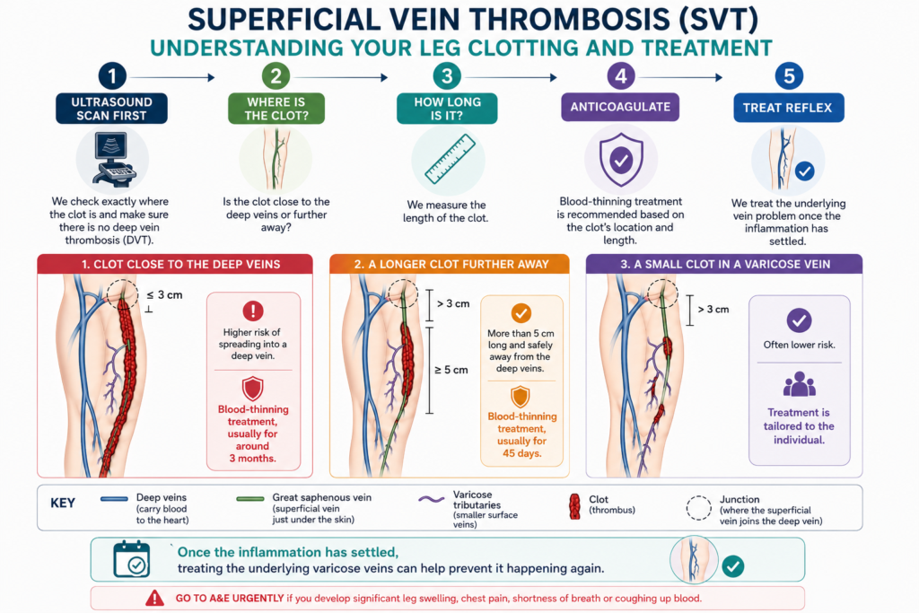
Patient infographic showing duplex assessment and treatment pathway for superficial vein thrombosis
How we assess and manage superficial vein thrombosis at Sheffield Vein Clinic.

This patient’s clot fell into the lower-risk “small clot in a varicose vein” category shown in the pathway above — which is why treatment was tailored specifically to what the scan showed, rather than a one-size-fits-all approach.

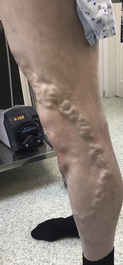
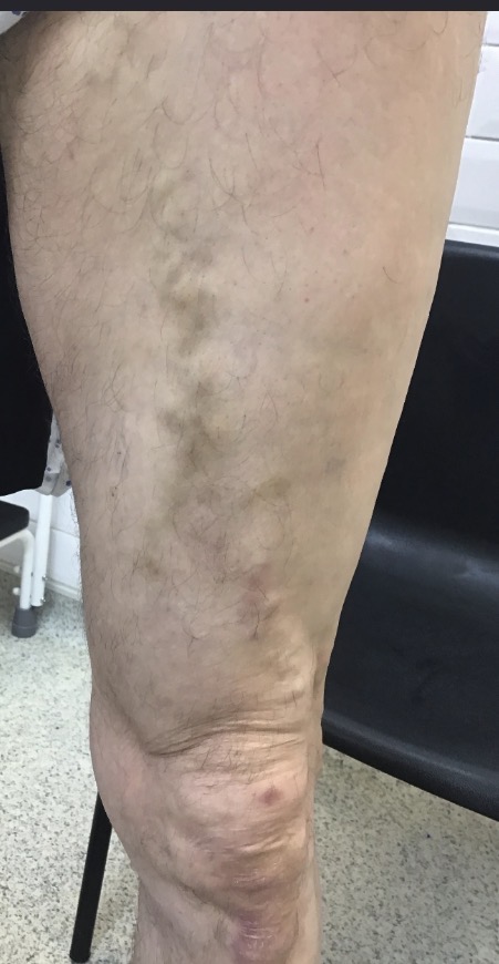
Close-up of the thrombophlebitic segment of the varicose vein, marked out ahead of treatment
Fig. 2 — A closer look at the inflamed segment, marked out ahead of treatment. The skin change follows the exact path of the vein underneath.

Getting the full picture with duplex ultrasound

A duplex ultrasound scan does two jobs at once: it shows us the structure of the vein (is there a clot, and how big is it?) and it shows us the blood flow (is it moving normally, or has it stopped in places?). For thrombophlebitis, the single most important measurement is the distance between the clot and the sapheno-femoral junction (SFJ), where the great saphenous vein joins the deep venous system. The closer the clot sits to that junction — or if it fills the vein all the way up to it — the more seriously it needs to be managed, sometimes with blood-thinning medication as a precaution.

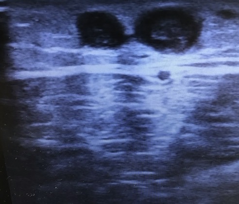
Duplex ultrasound scan of the affected varicose vein used to confirm the superficial clot
Fig. 3a — Ultrasound scan of the affected vein, used to confirm the clot and check how far it extends.
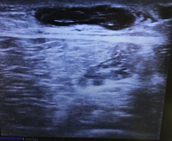
Second duplex ultrasound view confirming the extent of the thrombus in the superficial vein
Fig. 3b — A second view of the same area, confirming the clot’s position relative to the junction with the deep veins.
What the scan told us

The clot was confined to the superficial varicosity, close enough to the sapheno-femoral junction (SFJ) that a short course of anticoagulation was the sensible, guideline-led choice rather than compression alone. Management followed the European Society for Vascular Surgery (ESVS) clinical practice guidelines on the management of superficial vein thrombosis.

Settling the clot before treating the vein

In line with ESVS guidance, we prescribed a short course of anticoagulation — apixaban 2.5mg twice daily for one month — alongside two weeks in a class 2 compression stocking. This combination calms the acute clot and inflammation while the stocking supports the vein and reduces swelling.

This reflects the management chosen for this specific patient, based on their individual duplex ultrasound findings. It is not generic prescribing advice: anticoagulation decisions depend on the clot’s position, its extent, duplex findings and individual clinical assessment, and should always be made following your own specialist review.

The patient was brought back for a repeat duplex ultrasound to confirm the clot hadn’t propagated towards the deep venous system and was resolving as expected. The rescan was reassuring: the thrombus was settling nicely, with no extension, and the patient’s symptoms had noticeably improved. With the acute phase safely behind us, definitive treatment of the underlying vein was booked for six weeks later.

Treating the cause, not just the symptom

Six weeks on, with the clot fully settled, the patient underwent an uncomplicated, unilateral procedure: endovenous laser ablation (EVLA) of the full length of the incompetent left great saphenous vein, combined with foam sclerotherapy of the remaining varicose tributaries — both performed under local anaesthetic and ultrasound guidance, in line with ESVS treatment guidelines. EVLA closes the faulty GSV from the inside using heat from a laser fibre, while foam sclerotherapy treats the smaller branch veins feeding off it. This step matters because the thrombophlebitis was a direct symptom of that underlying GSV reflux. Treating the clot alone, without closing the reflux driving it, would have left the door open for the same thing to happen again somewhere along the vein’s length.

Recovery

At follow-up, the result was excellent. The area that had been red, hard, and painful was soft and comfortable again, with only some fading brown discolouration left behind — a normal, temporary mark from the original clot as the body reabsorbs it, which continues to lighten over the following months. The vein itself was no longer the bulging, engorged structure it had been, the procedure was uncomplicated throughout, and the patient was delighted with the outcome. You can see more examples of results like this in our before & after gallery.

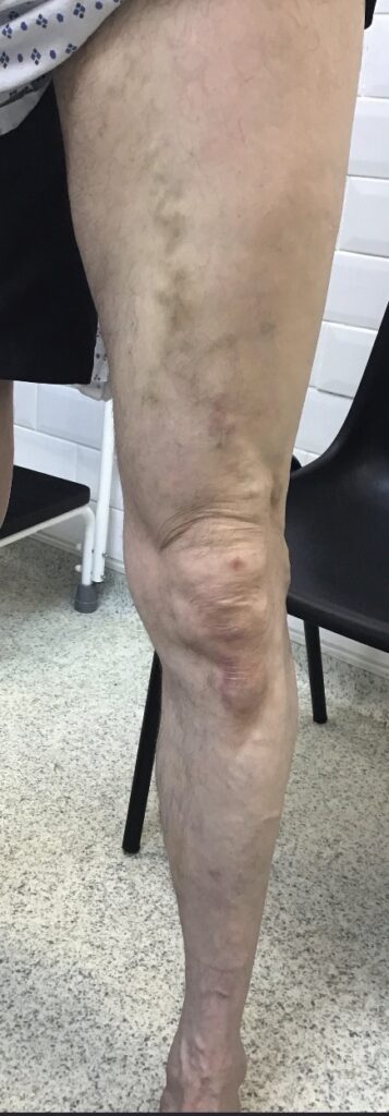
Before
Varicose vein with thrombophlebitis before EVLA treatment
Before EVLA — the varicose vein prior to treatment.
After
Leg following EVLA treatment, follow-up view showing resolved thrombophlebitis and vein
After EVLA — early follow-up. The inflammation has settled and the vein is no longer prominent.
After (later follow-up)
Later follow-up view after EVLA treatment showing the leg almost fully healed
After EVLA — later follow-up, nearly fully healed.

The takeaway

  • A red, hard, painful lump on a varicose vein is not “just a bruise” — get it scanned.
  • Duplex ultrasound is quick, painless, and answers the question that actually matters: has the clot reached the deep veins?
  • Thrombophlebitis is often a sign of an underlying vein that needs treating, not just the clot itself.
  • Treating the source vein with EVLA gives a lasting result and reduces the chance of it happening again.

Frequently asked questions

Is a hard, red lump on a varicose vein serious?

It should always be checked. It’s usually superficial thrombophlebitis — uncomfortable, but not immediately dangerous. The important step is a duplex ultrasound to check the clot hasn’t spread towards the deep veins.

What is the difference between thrombophlebitis and a DVT?

Thrombophlebitis affects a superficial vein near the skin and is visible as redness and a firm lump. A DVT affects the deeper veins, doesn’t show on the surface, and carries a higher risk of complications. Superficial thrombophlebitis is generally the less risky of the two, but a clot can still propagate, dislodge, or — in the highest-risk pattern — fill the entire great saphenous vein and reach the sapheno-femoral junction, putting it at the doorway to the deep system. In rare cases a fragment can travel to the lungs (pulmonary embolism). A duplex scan tells the two apart and identifies this risk.

Do you always need blood thinners for thrombophlebitis?

Not always — it depends how close the clot sits to the junction with the deep venous system, which the scan measures. In this case, the clot’s position meant a short course of apixaban plus a class 2 compression stocking was the right, guideline-led approach; clots further from the junction can sometimes be managed with compression alone. This decision is always individual and depends on your own scan findings.

Will thrombophlebitis come back if I don’t treat the varicose vein itself?

It can. Thrombophlebitis is often a sign of an underlying faulty vein. Treating that vein directly, for example with EVLA, addresses the cause and lowers the chance of a repeat episode.

How long does the discolouration take to fade?

The brownish staining comes from iron deposits left behind as the clot resolves. It typically fades gradually over several months and isn’t a sign of an ongoing problem.

Concerned about a painful varicose vein?

If a varicose vein has suddenly become hard, tender or inflamed, a specialist assessment and duplex ultrasound can establish what is happening and whether further treatment is needed.

Request a Vein Assessment

This case is shared for educational purposes with patient consent. Identifying details have been removed or altered to protect patient confidentiality. Individual treatment decisions depend on clinical assessment and duplex ultrasound findings.

About the author

Professor Stephen Goode is a Consultant Interventional Radiologist and lead clinician at Sheffield Vein Clinic.