772–774 ECCLESALL ROAD · SHEFFIELD · S11 8TB info@sheffieldveinclinic.co.uk
Real Case · Complex Varicose Veins
Most people picture varicose veins on the calf or behind the knee. It’s far less well known that they can track all the way down into the foot itself, where they become some of the hardest veins in the body to treat safely. Here’s a real case from our clinic, from the first consultation through to an eight-week follow-up.
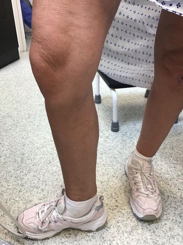
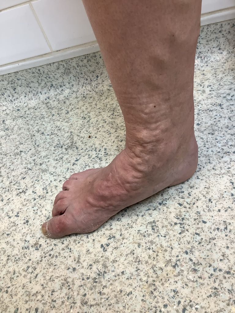
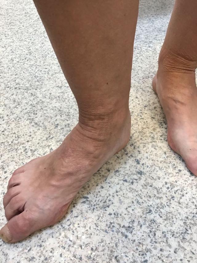
This patient, a 56-year-old woman, came to us with large, dilated, recurrent varicose veins on both legs, despite open surgical vein surgery under general anaesthetic some 20 years earlier. The veins had come back, and on the right side had progressed further than most: a complex tangle of varicosities extended all the way down into the foot itself. They were a major cosmetic concern, and by the end of the day they ached and throbbed enough to genuinely affect her quality of life.
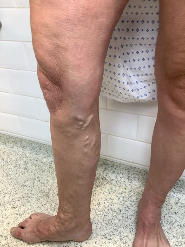
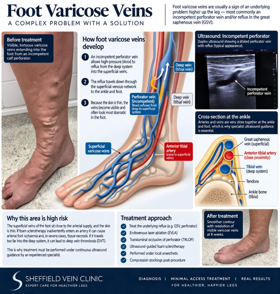
Varicose veins in the foot are almost always a sign of a problem further up the leg, not a problem that starts in the foot. Superficial leg veins are fed by “perforator” veins — short connecting vessels that pass through the muscle to link the surface veins with the deep venous system. When a perforator becomes incompetent, it allows high-pressure blood to reflux the wrong way, down through the superficial network, all the way to its lowest point: the ankle and foot. Because the skin there is thin, this is often where the disease looks its most dramatic, even though the fault lies higher up.
Not in terms of danger to health — but it is more serious in terms of difficulty to treat well. The foot’s superficial veins sit close to its arterial supply, and the skin covering them is thin and mobile. That proximity is what makes this one of the more technically demanding corners of venous surgery, and it’s why foot varicosities are often left untreated by clinics that don’t have experience working this low down the leg.
A recurrence like this usually announces itself fairly clearly, but it’s worth knowing what to look for after previous vein treatment:
Treating varicose veins at the ankle and foot carries risks that don’t apply higher up the leg. If foam sclerotherapy inadvertently enters an artery instead of a vein, it can cause arterial foot ischaemia and, in severe cases, tissue necrosis. If it travels too far into the deep system at the ankle, it raises the risk of deep vein thrombosis (DVT). This is exactly why cases like this are planned and executed under continuous ultrasound guidance, by someone comfortable reading both the venous and arterial anatomy of the ankle and foot in real time.
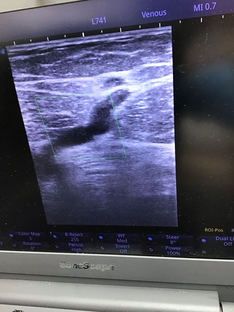
Duplex ultrasound scanning did the same two jobs it always does: mapping the structure of the veins, and confirming which way the blood was actually flowing. For a recurrent case like this, the key question is which vessel is now driving the disease, since the original feeding source may already have been dealt with years earlier.
Residual reflux was confirmed in both great saphenous veins (GSV), and — specifically driving the right-sided foot involvement — a large, dilated, incompetent right calf perforator vein. This wasn’t one problem but two feeding vessels that both needed closing: the GSV reflux on both sides, and the calf perforator on the right.
The plan addressed both feeding sources directly, using minimally invasive, keyhole techniques under local anaesthetic throughout. Bilateral endovenous laser ablation (EVLA) of the recurrent GSV was performed via ultrasound-guided Seldinger access with tumescent (1% lidocaine) anaesthesia around the vein before firing the laser, closing the main superficial trunk driving reflux on both sides.
TRLOP of the right calf perforator is a more delicate procedure: the incompetent perforator is accessed directly using an 18-gauge needle under ultrasound guidance, the needle is exchanged for a laser fibre positioned precisely inside the perforating vein, and a short, focused segment of around 1–2cm is ablated at 8.5 watts, carefully staying clear of the adjacent deep vein. TRLOP achieves in a keyhole procedure what open surgery would otherwise need a formal incision and general anaesthetic to attempt, without the difficulty of locating and tying off the vessel by hand.
Foam sclerotherapy (1%) was then used on the residual varicosities on both legs, accessed from above and allowed to reflux down into the diseased foot veins under direct, continuous ultrasound monitoring, watching closely for any sign of the foam tracking too far toward the deep system or straying into arterial territory. No complications were encountered, and the procedure was completed successfully throughout.
Class 2 compression stockings were worn for two weeks after treatment to support healing. Patients receive a post-operative information sheet covering what to expect, a structured telephone consultation with the clinical vein specialist team at one week, and a direct line to the team for anything unexpected in between.
At eight-week follow-up, repeat duplex ultrasound confirmed the right calf perforator was fully occluded, and both the leg and the foot varicosities it had been feeding had resolved. The result was an excellent technical and symptomatic outcome, and the patient was especially pleased that the foot varicosities, her main cosmetic concern from the outset, had resolved along with everything else.
At six months post-op, the patient continues to feel the benefit: durable symptomatic relief, a lasting cosmetic improvement, and, by her own account, a significant boost in confidence. You can see more examples of results like this in our before & after gallery.
Can varicose veins really extend into the foot?
Yes. It’s less common than calf or thigh involvement, but when an incompetent perforator or trunk vein sits low enough in the leg, reflux can track all the way down to the ankle and foot, where the thin skin often makes the veins look especially prominent.
Why is treating foot veins riskier than treating veins higher up the leg?
The foot’s superficial veins sit close to its arterial supply. Treatments like foam sclerotherapy have to be watched under continuous ultrasound to make sure they stay in the venous system — straying into an artery can cause serious complications, including tissue damage.
What is TRLOP, and how is it different from traditional perforator surgery?
TRLOP (TransLuminal Occlusion of Perforator) closes an incompetent perforator vein from the inside using a laser fibre placed directly into it under ultrasound guidance. It’s a keyhole alternative to open surgical ligation, which requires a formal incision, general anaesthetic, and can be difficult to precisely locate the vessel.
I’ve had varicose vein surgery before and they’ve come back — why does this happen?
Recurrence is common, particularly with older open surgical techniques, if some of the underlying reflux isn’t fully addressed at the time. A fresh duplex ultrasound can usually identify exactly which vessel is now driving the recurrence, as it did here with the calf perforator.
How long is recovery after this kind of combined procedure?
Most patients wear a class 2 compression stocking for around two weeks and return to normal activity quickly. These are walk-in, walk-out procedures done under local anaesthetic, without the extended recovery associated with open surgery.
Don’t assume it’s untreatable because of where it is. A duplex ultrasound can identify the source and map out a plan.
Book a Duplex AssessmentThis case is shared for educational purposes with patient consent. Identifying details have been removed or altered to protect patient confidentiality.
About the author
Professor Stephen Goode is a Consultant Interventional Radiologist and Professor and Research Lead at Sheffield Teaching Hospitals NHS Trust, and lead clinician at Sheffield Vein Clinic.