Real Case · Posterior Thigh Perforator & Varicose Veins
Back on the Tools: Treating a Large Posterior Thigh Perforator in a Young Construction Worker
Varicose veins have a reputation as an older person’s problem — something cosmetic, something that can wait. This case shows why that reputation is wrong. Our patient was 32, worked full-time in construction, and had spent five years watching his symptoms get steadily worse until they were affecting his ability to do his job. Here’s how we found the cause, treated it without a single incision, and got him back to work.
Clinical presentation
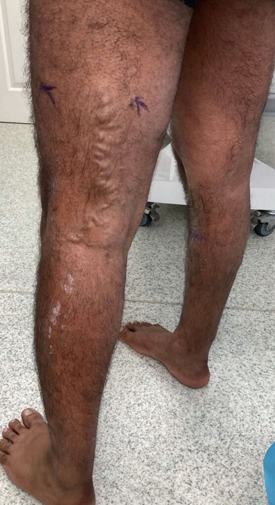
This patient came to us with large, dilated varicose veins on both legs that had been worsening for around five years. They were a significant cosmetic concern, but far from just cosmetic: by the end of a working day he had real pain and throbbing, and the skin on both legs had started to show haemosiderin staining — a brownish discolouration that’s a marker of longstanding, more advanced venous insufficiency, not something we’d typically expect to see at 32. Being on his feet on site all day made the symptoms particularly hard to manage, and the veins were starting to affect his working life directly.
What is a perforator vein, and why does it matter?
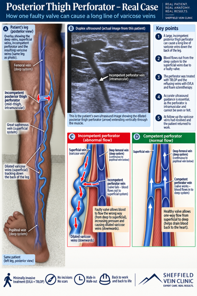
Perforator veins are the short connecting veins that link the superficial venous system (just under the skin) to the deep venous system (which does most of the work returning blood to the heart). Normally, one-way valves inside them keep blood flowing in a single direction — from superficial to deep. When a perforator’s valve fails, blood is pushed the wrong way, back out into the superficial veins — a pathological outward, or refluxing, flow — adding extra pressure and volume on top of whatever reflux is already present in veins like the GSV. A large, incompetent perforator can be the main engine driving a stubborn or recurrent pattern of varicose veins, which is exactly what duplex ultrasound was used to confirm here.
Deep vein, perforator and superficial veins: how they connect
Duplex ultrasound findings
Duplex ultrasound scanning showed bilateral great saphenous vein (GSV) reflux, and — of particular note — a large, dilated, incompetent perforating vein deep in the left posterior thigh. This perforator was intramuscular, sitting well below the surface, which is part of what makes these veins technically demanding to treat: you’re working with direct, image-guided access into a vessel you can’t see or feel, using ultrasound as your only eyes.
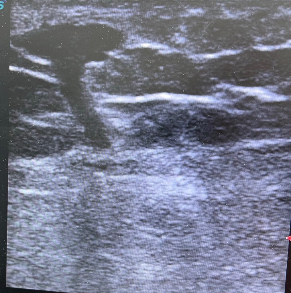
Based on the scan findings, the treatment plan was bilateral endovenous laser ablation (EVLA) of the full length of both GSVs, TRansLuminal Occlusion of Perforator (TRLOP) of the left posterior thigh perforator, and foam sclerotherapy to the remaining varicose tributaries on both legs — all performed through keyhole access under ultrasound guidance, with no incisions and no scarring.
Why this case was unusual
This case is a good illustration that varicose veins aren’t always driven by conventional great or small saphenous vein reflux alone. Duplex ultrasound identified a large, incompetent perforating vein deep in the left posterior thigh — an intramuscular vessel, sitting well below the surface, allowing pathological outward reflux flow from the deep venous system into the superficial varicose veins on top of the bilateral GSV reflux already present. Recognising and treating the actual source of reflux, rather than just the visible surface veins, is what a thorough duplex assessment made possible here. It’s also a reminder that this isn’t purely a condition of older age: our patient was only 32, and the impact on a physically demanding job was real.
Treatment
Post-operative DVT is one of the main risks we plan around in any venous procedure, so every patient undergoes a dedicated DVT risk assessment at their initial consultation, with a tailored thromboprophylaxis protocol set in advance. On the day, a further duplex scan is used to mark up the exact access points for the endovenous laser — for a TRLOP procedure in particular, precise pre-operative planning is essential, since the target is a dilated, refluxing perforator sitting within the muscle rather than just under the skin. A WHO checklist and safety stop is completed before starting, and the legs are prepped with 1% chlorhexidine using aseptic technique. Everything is done under local anaesthetic: targeted, ultrasound-guided local anaesthetic injection means there’s no need for a general anaesthetic at all, which is what allows this to be a genuine walk-in, walk-out treatment.
Access to the GSV is gained under ultrasound guidance using Seldinger catheter and wire-exchange technique, with the laser fibre advanced to the level of the sapheno-femoral junction. Tumescent anaesthesia (1% lidocaine) is instilled around the vein and the fibre before the laser is fired, ablating the GSV under direct ultrasound control.
The TRLOP procedure is the more technically demanding part of this case. Direct access into the dilated perforating vein is achieved with an 18-gauge Seldinger needle under ultrasound guidance, after local anaesthetic to the access site. The needle is then exchanged for the laser probe, which is placed directly into the perforator — with real care taken to keep it clear of the adjacent deep femoral vein. Tumescent anaesthesia is instilled around the laser under ultrasound guidance, and ablation is performed over a focused 1–2 cm segment at 8.5 watts, delivering 135 joules. Foam sclerotherapy (3% solution) is then used to treat the remaining varicose tributaries. TRLOP gives a minimally invasive, precisely targeted way of closing an incompetent deep perforator. The alternative — open surgical ligation — means a formal incision, a general anaesthetic, and can be genuinely difficult surgery, since finding and isolating the perforator by hand is not straightforward.
The main risk with perforator ablation is inadvertent damage to the adjacent deep vein, which can lead to DVT — this is why ultrasound guidance and careful probe positioning throughout the TRLOP procedure are so important. Other recognised risks include nerve injury, bleeding, thrombophlebitis, and skin staining. In this case, no complications were encountered and the procedure was completed with full technical success.
Recovery and follow-up
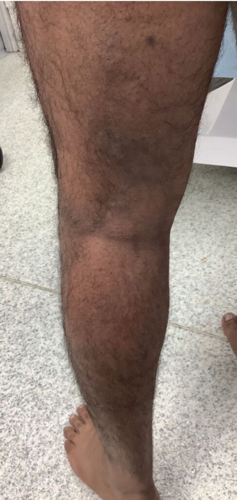
Class 2 compression stockings were worn for two weeks after the procedure to support healing and reduce the risk of complications. Patients receive written information covering common post-operative issues, a telephone follow-up with the clinical vein specialist team at one week, and a dedicated contact number for anything that needs urgent attention — with a direct route back to the treating consultant if needed. At eight-week review, the perforator was fully occluded on repeat duplex scanning, along with resolution of the associated varicose veins. The patient reported a significant improvement in his symptoms and was already working more comfortably, with no pain by the end of the day. A small area of residual bruising was drained at this visit, a touch-up foam sclerotherapy treatment was given to a few remaining tributaries, and compression stockings were reapplied for a further week.
Before and after
Result
At six months, the result has held: full symptomatic relief, a markedly improved cosmetic appearance, and a patient who’s back to full, pain-free working days on site. In his own words, it’s “as though nothing has been done” — he’s simply back to normal life, which for a physically demanding job is exactly the outcome that matters most.
This case is a reminder that varicose veins are not purely a condition of older age, and not purely cosmetic. Left untreated, they can meaningfully affect a young, physically active person’s ability to work. It’s also a good example of how far endovenous techniques have moved on from traditional open surgery — from large incisions, general anaesthesia, and a long recovery, to a targeted, walk-in walk-out procedure performed entirely under local anaesthetic and ultrasound guidance, in line with NICE guideline CG168. You can read more real patient outcomes like this in our clinical case reports, or see further examples in our before & after gallery.
Key learning points
- Varicose veins can be seriously symptomatic — and disabling for physically demanding work — even in patients in their 30s.
- A large, incompetent perforator vein can be the hidden driver behind a stubborn pattern of varicosities, and duplex ultrasound is what finds it.
- TRLOP allows a deep, intramuscular perforator to be closed through a single needle access point, without the incision and general anaesthetic that open surgical ligation would require.
- Treating the GSV reflux and the perforator together, rather than just the visible surface veins, gives a durable result and reduces the chance of recurrence.
Frequently asked questions
Are varicose veins just a cosmetic issue?
Not necessarily. As this case shows, they can cause real daily pain and throbbing, and in longstanding cases can lead to skin changes such as haemosiderin staining — a sign the venous insufficiency is becoming more advanced, regardless of the patient’s age.
What is a perforator vein, and why does it need separate treatment?
Perforator veins connect the superficial and deep venous systems. When their valves fail, they push blood the wrong way and can drive a pattern of varicose veins on their own. Duplex ultrasound identifies them, and a large, refluxing perforator like this one needs to be closed directly — treating the GSV alone would not have addressed it.
How is TRLOP different from traditional perforator surgery?
Traditional surgical ligation of a perforator vein requires an incision and general anaesthetic, and can be difficult surgery to localise the vessel accurately. TRLOP instead uses ultrasound-guided needle access and a laser fibre to close the perforator from the inside, under local anaesthetic, with no incision.
Is this a walk-in, walk-out procedure?
Yes. All of the treatment in this case — bilateral GSV ablation, TRLOP of the perforator, and foam sclerotherapy — was performed under local anaesthetic in a single visit, with the patient walking in and walking out the same day.
Concerned about varicose veins affecting your work or daily life?
A duplex ultrasound assessment identifies exactly what’s driving your symptoms — including perforator veins that aren’t visible on the surface.
Book a Duplex AssessmentThis case is shared for educational purposes with patient consent. Identifying details have been removed or altered to protect patient confidentiality. Treatment followed NICE clinical guideline CG168 on the diagnosis and management of varicose veins.
About the author
Professor Stephen Goode is a Consultant Interventional Radiologist and Professor and Research Lead at Sheffield Teaching Hospitals NHS Trust, and lead clinician at Sheffield Vein Clinic.